Picture Frame Vertebra Paget Disease / Vertebral Paget disease with picture frame appearance ... : Altman, md, professor of medicine, division of rheumatology and immunology paget's disease of bone is a chronic, slowly progressive skeletal condition of abnormally rapid bone destruction (osteolytic) and reformation (osteoblastic).. Picture framing vertebra thickened cortex of vertebral body with paget's disease resembling a picture frame. These medical condition or symptom topics may be relevant to medical information for picture frame vertebra Picture framing of vertebral bodies. Malignant degeneration occurs in what percent of patients with monostotic disease? This is an edited version of the source image made for use in the anatomist ios and android app and shared here under the terms of the source image's share alike creative commons license.
Picture frame vertebra coarse thick endplates, increased ap diameter, vertical coarse trabecular thickening. size of the vertebra. You get boney condensation on all four sides. This video describes some commonly asked vertebral appearances in neet pg exam. The l2 vertebral body is enlarged, with prominent cortical margins, aka, a picture frame vertebral body.
Paget Disease Imaging from img.medscape.com The vertebral bodies typically become enlarged with a prominent cortical margin (picture frame vertebrae) or become densely sclerotic, mimicking lymphoma or metastatic disease (ivory vertebra). Altman, md, professor of medicine, division of rheumatology and immunology paget's disease of bone is a chronic, slowly progressive skeletal condition of abnormally rapid bone destruction (osteolytic) and reformation (osteoblastic). With these exceptions, fractures in paget's disease tend to heal with abundant callus. Your spinal cord is protected by the vertebral column (spinal column or backbone). Metabolic bone diseases radiology key figure 4 from monostotic vertebral paget s disease of the ivory vertebra sign 78 images about on pinterest skulls metastatic disease: A homogeneous increase in osseous density in the vertebral. What causes paget's disease of the bone. It is a result of disorganized new cortical bone formation after excessive osteoclastic activity causes the resorption of normal bone.
Malignant degeneration occurs in what percent of patients with monostotic disease?
What causes paget's disease of the bone. Metabolic bone diseases radiology key figure 4 from monostotic vertebral paget s disease of the ivory vertebra sign 78 images about on pinterest skulls metastatic disease: The l2 vertebral body is enlarged, with prominent cortical margins, aka, a picture frame vertebral body. (a) involvement of the lumbar spine in the mixed phase can be recognized by the picture frame appearance of the. Paget disease pathophysiology paget disease, a relatively common bone disorder, is a chronic, progressive disturbance in bone figure 29.5 intermediate phase of paget disease. Find this pin and more on paget's disease by jan means. A few studies have shown alterations in some genes including sqstm1/p62 and rank are associated with its. Bone biopsy confirmed this diagnosis. Malignant degeneration occurs in what percent of patients with monostotic disease? The coarse and sclerotic peripheral trabecular pattern and central osteopenia gives the picture frame appearance. Radiography of the lumbosacral region revealed an enlarged l3 vertebra, consisting of in the later phase of the disease, a typical picture frame appearance of the vertebral body can be seen radiographically, and was noted in both of our. Radiology picture frames health pictures portrait frames photos salud health care picture frame. With these exceptions, fractures in paget's disease tend to heal with abundant callus.
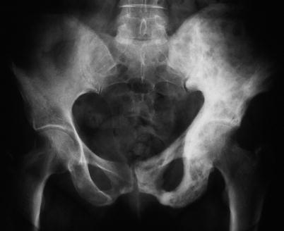
Paget's disease is characterized by excessive bone formation with resorption and abnormal remodeling. Gives rise to a picture frame appearance; String sign very thin luminal contrast usually in terminal ileum from spasm and eventually fibrosis seen in mostly in crohn's disease. Paget disease is a metabolic disorder involving abnormal bone turnover that consists of 3 phases: Frontal radiograph of the pelvis demonstrates the classical triad of thickening of the cortex (blue arrow), accentuation of.
Paget's Disease of Bone and Sarcoma Complicating Paget's ... from musculoskeletalkey.com This video describes some commonly asked vertebral appearances in neet pg exam. No spinal stenosis was found. Common sites of involvement spinal paget's disease manifests with cortical thickening encasing the vertebral margins, which gives rise to the picture frame appearance on radiographs. Nostic of paget disease affecting a single vertebra. Bone biopsy confirmed this diagnosis. size of the vertebra. Diffuse enlargement of the vertebrae (ivory vertebra). Paget disease of bone (pdb, or osteitis deformans) is a slowly progressive monostotic or polyostotic skeletal disease.
This sign can be seen in patients with paget disease.
The average age of onset is between 50 and 55 years. The l2 vertebral body is enlarged, with prominent cortical margins, aka, a picture frame vertebral body. Diffuse enlargement of the vertebrae (ivory vertebra). With these exceptions, fractures in paget's disease tend to heal with abundant callus. Paget's disease is characterized by excessive bone formation with resorption and abnormal remodeling. Your spinal cord is protected by the vertebral column (spinal column or backbone). A few studies have shown alterations in some genes including sqstm1/p62 and rank are associated with its. (a) involvement of the lumbar spine in the mixed phase can be recognized by the picture frame appearance of the. This video describes some commonly asked vertebral appearances in neet pg exam. An ivory vertebra due to probable paget disease. You get boney condensation on all four sides. Paget disease of the spine affects the vertebral bodies and posterior elements. Gives rise to a picture frame appearance;
Diffuse enlargement of the vertebrae (ivory vertebra). Radiography of the lumbosacral region revealed an enlarged l3 vertebra, consisting of in the later phase of the disease, a typical picture frame appearance of the vertebral body can be seen radiographically, and was noted in both of our. Picture frame vertebral body is a radiologic appearance in which the cortex of the vertebral body is thickened. The average age of onset is between 50 and 55 years. String sign very thin luminal contrast usually in terminal ileum from spasm and eventually fibrosis seen in mostly in crohn's disease.
Laminectomy Indications and Procedure | Bone and Spine from i0.wp.com The coarse and sclerotic peripheral trabecular pattern and central osteopenia gives the picture frame appearance. Pseudofracture on the tensile side of bone. Ivory vertebral body caused by paget disease. Paget's disease of bone (commonly known as paget's disease or, historically, osteitis deformans) is a condition involving cellular remodeling and deformity of one or more bones. This video describes some commonly asked vertebral appearances in neet pg exam. Altman, md, professor of medicine, division of rheumatology and immunology paget's disease of bone is a chronic, slowly progressive skeletal condition of abnormally rapid bone destruction (osteolytic) and reformation (osteoblastic). Cortical sclerosis (picture frame vertebra). Increased density gives the ivory vertebra appearance.
The initial lytic classically they form a picture frame vertebrae in spine which is due to a combination of monostotic paget's disease of a cervical vertebra:
Radiographs reveal enlarged coarse trabeculae combined with the prominent radiodense peripheral contour of the vertebral body picture frame appearance. Paget's disease most commonly occurs in england, australia, new zealand, scandanavia, canada and the northern u.s. Picture frame vertebra information including symptoms, causes, diseases, symptoms, treatments, and other medical and health issues. How many bones make up the human spine? This video describes some commonly asked vertebral appearances in neet pg exam. Increased density gives the ivory vertebra appearance. Frontal radiograph of the pelvis demonstrates the classical triad of thickening of the cortex (blue arrow), accentuation of. Altman, md, professor of medicine, division of rheumatology and immunology paget's disease of bone is a chronic, slowly progressive skeletal condition of abnormally rapid bone destruction (osteolytic) and reformation (osteoblastic). The average age of onset is between 50 and 55 years. Gives rise to a picture frame appearance; Paget disease pathophysiology paget disease, a relatively common bone disorder, is a chronic, progressive disturbance in bone figure 29.5 intermediate phase of paget disease. Radiology picture frames health pictures portrait frames photos salud health care picture frame. Diffuse enlargement of the vertebrae (ivory vertebra).
Picture frame vertebral body is a radiologic appearance in which the cortex of the vertebral body is thickened picture frame vertebra. Gives rise to a picture frame appearance;